
The Next Generation of Specimen Imaging for the Gross Room.
In today’s fast-paced pathology lab, PAs need X-ray imaging in the gross room to reduce turnaround time and eliminate specimen transport to radiology or mammography, while enabling accurate identification of clips, markers, and microcalifications.
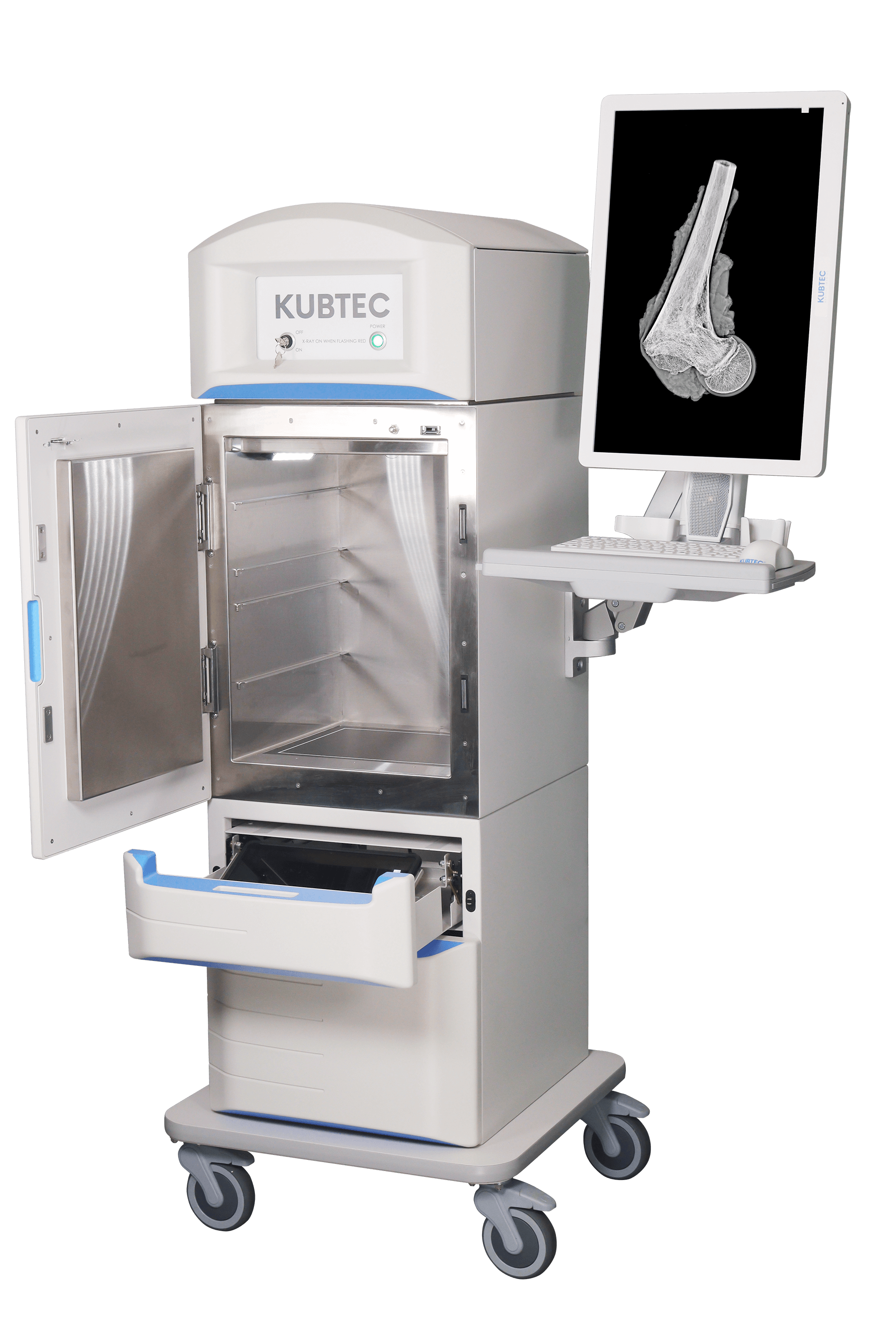
The PICASSO Plus System redefines imaging in the Pathology Lab. It includes workflow features designed specifically for the gross room as well as being the only Pathology specimen imaging system featuring Amorphous Selenium Direct Capture Technology. So the high image quality used to diagnose breast cancer can now also be used to help analyze breast tissue in the pathology lab.
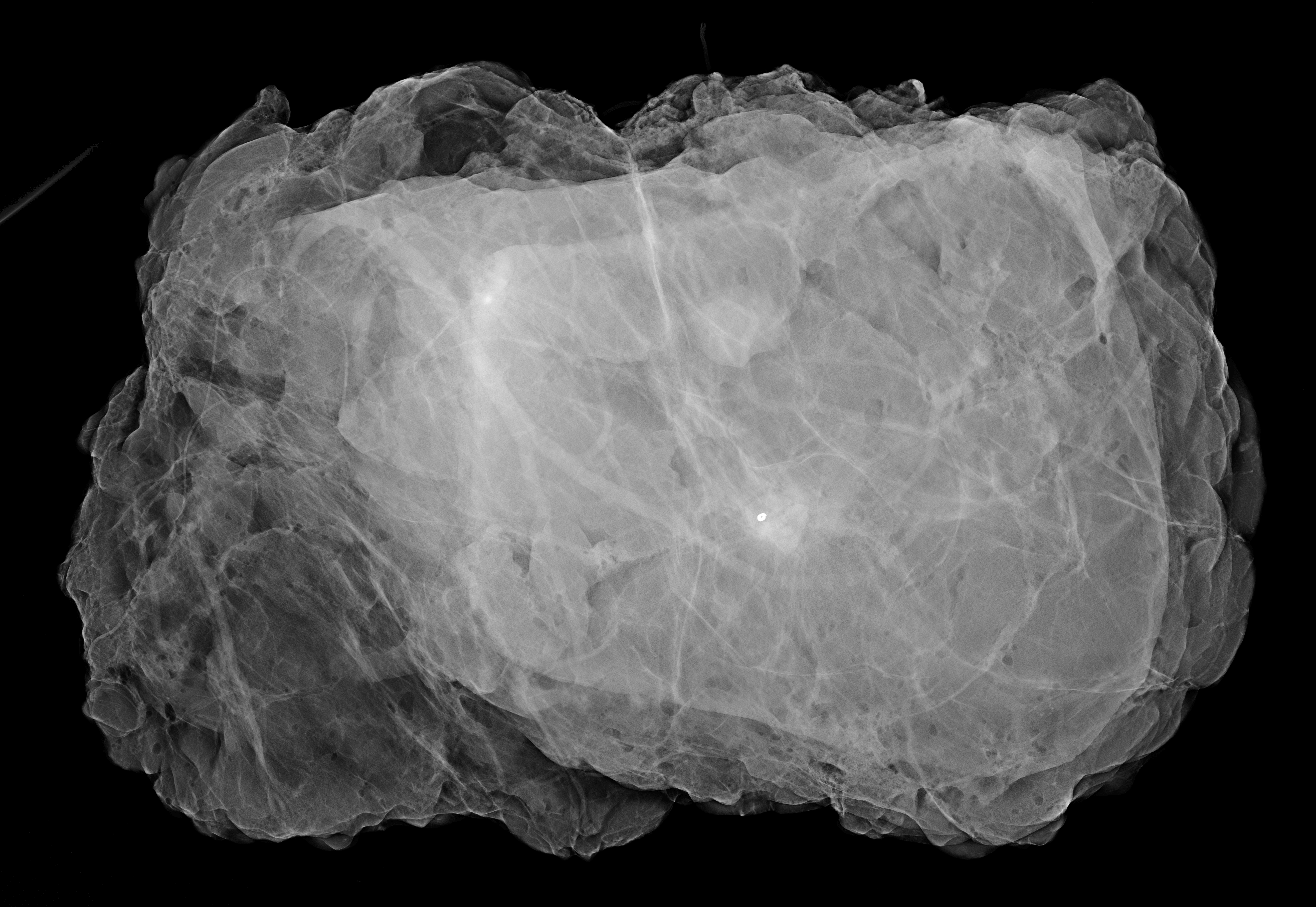
Blend both the X-ray and optical images to quickly locate surgical clips, margins, and microcalcifications to best gross specimens and select tissue to place into cassettes.
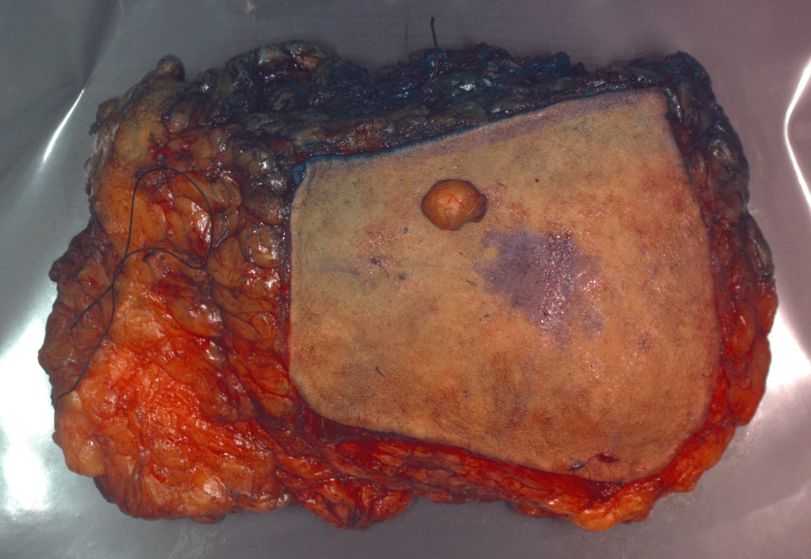
Creates optical images, enabling you to visually orient your specimen accurately, in real-time.

Combine X-ray and Optical Images for the Perfect Blend!

A 10” x 12” detector for larger specimens, like mastectomies.

Convenient access to images and annotation tools anywhere in the gross room.

Export your X-ray and optical images directly to LIS.

Innovative speech recognition technology for true hands-free navigation

A graphical representation to distinguish between calcified and decalcified bone.

Notifies you if a specimen is accidentally left inside the system.

Operate the optional Pablo™ Tablet without touching it.


Contact us to see The PICASSO® Plus Specimen Radiography System in action.

